Substages of Prophase I
Stages of Meiosis
EUGENE M. MCCARTHY, PHD GENETICS
| Online Biology Dictionary |
Prophase I is by far the most complicated phase of meiosis. It is also much longer in meiosis than in mitosis. During this stage, homologs join (synapse) along their lengths and exchange DNA. Prophase I is itself divided into the five substages explained and diagrammed below.
|
| Leptotene |
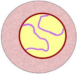
In this first substage of prophase I, the chromosomes have appeared within the nuclear envelope (shown in the diagram at right as a tan circle with a brown border), but are not yet fully condensed. In the diagram the two chromosomes of paternal origin are indicated in red, those of maternal origin, in blue. Each is a thin thread of DNA (lepto- is Greek for thin and -tene is Greek for ribbon or band) along which clearly defined beads of local coiling (chromomeres) can be seen. The chromosomes, while they have this threadlike form, are called chromatonemata (sing. chromonema; -nema is Greek for thread). The chromosomes appear single because the sister chromatids are still so tightly bound to each other that they cannot be separately seen. During this stage both telomeres of each chromosome are turned toward, and probably attached to, the same region of the nuclear envelope. Leptotene is also known as
-
leptonema;
and as
- the bouquet stage because all the telomeres tend to contact the nuclear envelope in one spot so that the looped chromosomes balloon out from that point like flower petals.
|
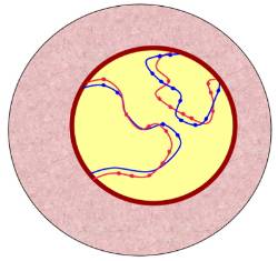
| Early Zygotene |
|
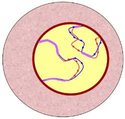
| Late Zygotene |
Zygotene
During zygotene homologs begin to unite (synapse) by coming into approximate alignment (zygo- is Greek for union, fusing, or yoking). Synapsis, the process of fusion that occurs between homologs begins at various points along the chromosome and extends outward, zipper-fashion, until complete. When synapsis is finished, the fused homologs look like a single chromosome under the light microscope, but they are actually double. The interface where two homologs unite, the synaptonemal complex, can be seen under an electron microcope.
In the diagram of early zygotene at right, the regions where the paternal and maternal homologs have fused is shown in purple. In the next diagram, representing late zygotene, both homolog pairs have fused over their entire lengths (so they are shown entirely in purple). Zygotene is also known as zygonema.
Tetrads: Once the homolog pairs synapse they are called tetrads (each has four chromatids; tetra is Greek for four) or bivalents. Bivalent is the preferred term, but tetrad is, nonetheless, the word more commonly used in most introductory biology classes. Bivalent is the better choice because there are equivalent names for other situations. For example, an unfused homolog is called a univalent. Three fused homologs, a common situation in plants, is called a trivalent, etc.
Pachytene
During pachytene the two sister chromatids of each chromosome separate from each other. This makes the chromosomes look thicker (pachy- is Greek for thick). Homologs are still paired at this point. Pachytene is also known as pachynema.
Crossing-over: Non-sister chromatids remain in contact throughout pachytene and a kind of localized breakage of the DNA occurs, which is followed by exchanges of DNA between them. This process is called crossing over. Crossing over produces "cross-over chromatids," each composed of distinct blocks of DNA, some blocks derived from the mother, others from the father.
Diplotene
At the beginning of this stage each chromatid of each chromosome is still fused to a chromatid of that chromosome's homolog (recall that sister chromatids are already separate at this point). As diplotene progresses, these initially fused non-sister chromatids begin to separate from each other. However, they cannot separate completely because they are still connected by two strands of DNA at each of the points where exchanges took place. At each such cross-over site, the two strands form an x-shaped structure called a chiasma (pl. chiasmata). The chiasmata then begin moving toward the ends of the chromatids. This process of sliding toward the ends is known as terminalization.
In oocytes, a special, extremely prolonged form of diplotene occurs called dictyotene. The primary oocyte undergoes the first three of the substages of prophase I (leptotene, zygotene, and pachytene) during late fetal life. The process is then suspended during diplotene until puberty or thereafter. Therefore, in dictyotene (and consequently prophase I) can last months or even years, depending on the type of organism in question. Diplotene is also known as diplonema.
Diakinesis
During this, the last stage of Prophase I, the nucleolus disappears, terminalization reaches completion, and the chromosomes coil tightly, and so become shorter and thicker. The nuclear envelope begins to disappear. The centrosomes reach the poles.