Metaphase I
Stages of Meiosis
|
|
EUGENE M. MCCARTHY, PHD GENETICS
| < | Mitosis | Meiosis | > |
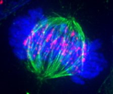 Photomicrograph of a metaphase cell. The massed
chromosomes (blue) can't be separately seen.
Photomicrograph of a metaphase cell. The massed
chromosomes (blue) can't be separately seen.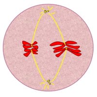 Simplified diagram of metaphase cell (compare with photo above).
Metaphase I
After prophase I, crossing-over is complete. The tetrads move to a plane — called the "metaphase plate" — halfway between the two poles of the cell. Next, the spindle fibers attach to the centromeres of each chromosome. Both kinetochores of each sister chromatid pair are turned toward the same pole. As a result, both kinetochores attach to spindle fibers from the same pole. This is a major difference between meiosis and mitosis. It causes the two members of each chromosome pair to be separated from each other during the next stage of meiosis, anaphase I.
Compare to metaphase of mitosis >>
Note: In metaphase of mitosis, the two kinetochores of each sister chromatid pair attach to spindle fibers from opposite poles. So each chromatid separates from its sister during the next step of mitosis, anaphase.