Gallbladder Anatomy
Online Biology Dictionary
|
|
|
|
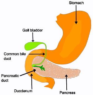
Basic gallbladder anatomy.
|
|
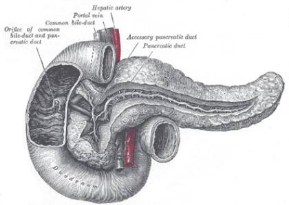
| Anatomy of the duodenum. |
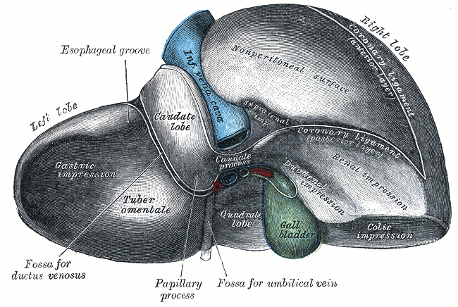
In human beings, the gallbladder is a small sac situated within the right side of the abdomen, on the under side of the liver, being attached to the right hepatic lobe. The Latin name for the gallbladder is the vesica fellea.
The gallbladder (which is pear-shaped, about 9 cm long and 2.5 cm in diameter at its widest) stores bile produced in the liver and discharges it through the cystic duct which joins with the hepatic ducts from the liver to form the common bile duct (ductus choledochus), which in turn connects with the duodenum, where the bile is discharged and aids digestion. The common bile duct unites with the pancreatic duct (duct of Wirsung) just before the two reach their common orifice, the papilla of Vater, in the duodenum.
Bile decreases the surface tension of water, so aiding in the emulsification of fats and oils, which is the action specifically of the bile salts (primarily sodium glycocholate and sodium taurocholate). The gallbladder contracts and discharges bile, when stimulated by the hormone cholecystokinin, which is produced by the duodenum in response to the presence of fatty foods.
| Note: Gall is the Anglo-Saxon word for bile, which is derived from Latin bilis. Gall is now generally used to refer only to the bile of animals, and not that of human beings. |
Most shared on Macroevolution.net:
Human Origins: Are we hybrids?
On the Origins of New Forms of Life
Mammalian Hybrids
Cat-rabbit Hybrids: Fact or fiction?
Famous Biologists
Dog-cow Hybrids
Georges Cuvier: A Biography
Prothero: A Rebuttal
Branches of Biology
Dog-fox Hybrids