Urinary Bladder Anatomy
Online Biology Dictionary
|
|
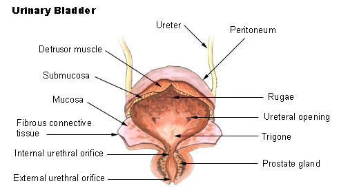
Major features of human urinary bladder anatomy
The urinary bladder ("YER-in-air-ee BLAH-der") is a muscular reservoir for urine, which it collects from the kidneys through the ureters. The Latin name for the urinary bladder is the vesica urinaria. It serves solely as a storage chamber and does not process or alter the urine in any way. The lower portion of the bladder forms a neck that continues as the urethra, through which urine is discharged from the body. Together with the urethra, the bladder forms a hollow, flexible, distensible bulb with a long neck.
In humans the bladder is situated in the pelvic cavity, lies anterior to the rectum in males, and anterior to the vagina and uterus in women. Its volume is about 0.25-0.50 liters, depending on the individual in question. "Retention" is an inability to empty the bladder. The ureterovesical valves prevent back flow of urine from the bladder into the ureters and are frequently the site where kidney stones become lodged ("impacted").
There is an interior lining of mucous epithelium, which is wrinkled in folds (rugae), that transitions to a layer of smooth muscle (the detrusor muscle, or detrusor urinae, which is enclosed in a fibrous layer of connective tissue. It is supported by several ligaments, one of which connects with the umbilicus. On its superior surface is a layer of peritoneum. The triangular area between the openings of the two ureters and the urethra is called the trigone.
A video about human urinary bladder anatomy:
Most shared on Macroevolution.net:
Human Origins: Are we hybrids?
On the Origins of New Forms of Life
Mammalian Hybrids
Cat-rabbit Hybrids: Fact or fiction?
Famous Biologists
Dog-cow Hybrids
Georges Cuvier: A Biography
Prothero: A Rebuttal
Branches of Biology
Dog-fox Hybrids