Human eye diagram
Online Biology Dictionary
|
|
 (Enlarge)
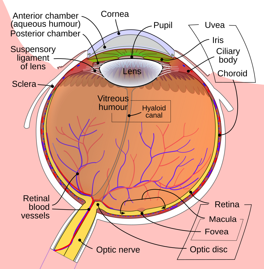
The human eye diagram displayed at right (click on it to see an enlarged image) shows all of the major features of human ocular anatomy. The eye has three layers or coats:
- the retina, which senses light;
- the uvea, which contains the nutritive blood vessels;
- the sclera and cornea, which form the protective coating.
The space between the lens and cornea, not labeled in the figure, is called the ocular chamber. It is filled with a watery fluid, the aqueous humor, and divided by the iris into an anterior and posterior chamber (the latter, which is hard to see in the figure, lies between the lens and iris). The main cavity of the eye is filled with the jelly-like vitreous humor. The optic disc, commonly known as the "blind spot," is the point where the optic nerve enters the back of the eye. The suspensory ligament, also known as the zonula ciliaris, holds the lens, while the ciliary muscles control the iris.
Online Biology Dictionary >>
Parts of the Eye:
- pupil: The opening through which light enters the eye.
- iris: The colored part of the eye that controls the amount of light passing through the pupil.
- retina: The light-sensitive tissue at the back of the eye. The retina converts light energy into nerve impulses, which are then sent to the brain via the optic nerve.
- lens: The clear structure behind the iris that focuses images on the retina.
- cornea: The clear outer covering at the front of the eye.
- macula: The small, sensitive area of the retina. The macula provides central vision.
- fovea: The central region of the macula. The fovea provides the clearest vision of any part of the retina.
- vitreous humor (or vitreous gel): The clear, jellylike substance that fills the eye.
Most shared on Macroevolution.net:
Human Origins: Are we hybrids?
On the Origins of New Forms of Life
Mammalian Hybrids
Cat-rabbit Hybrids: Fact or fiction?
Famous Biologists
Dog-cow Hybrids
Georges Cuvier: A Biography
Prothero: A Rebuttal
Branches of Biology
Dog-fox Hybrids