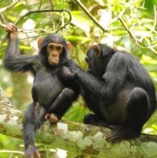
Low cancer rates in nonhuman primates
Humans differ markedly from nonhuman primates with respect to susceptibility to cancer. Thus, Puente et al. (2006) note that the the incidence of cancer in the latter is very low. They go on to say that
And this difference is not an artifact of small sample sizes. Beniashvili (1989, p. 423) comments that “Although tumors occur commonly in man, they have been rather infrequently reported in monkeys and apes, despite the fact that many thousands have been maintained in zoological gardens, primate centers, other research centers, and poliomyelitis vaccine production laboratories.”
Varki (2000) confirms these findings:
These rates of incidence do not seem to be explicable in terms of the shorter lifespans of nonhuman primates in comparison with humans. Thus, with regard to prostate cancer, Waters et al. (1998) comment that, "in the last decade, large numbers of elderly male nonhuman primates (last 10% of estimated lifespan) have been carefully evaluated; the absence of prostatic lesions confirms that prostate carcinoma is almost nonexistent in nonhuman primates.” And yet, in many regions of the world, prostate cancer is the most commonly diagnosed cancer among men (Center et al. 2012). Waters et al. go on to say nonhuman primates have very low rates of even benign prostatic lesions.
Siebold and Wolf (1973, p. 536, Table 2) reported on 1066 necropsies of nonhuman primates. Among these were 52 chimpanzees in which 6 tumors were observed, none of which were malignant. In the whole study, among all animals, they found only 24 tumors of any kind, just 4 of which were malignant, corresponding to a cancer incidence of 0.38 percent (compare this with the human death rate from malignant epithelial tumors alone of more than 20%). Stanford (2018) states that “Some of the most prevalent human diseases that occur later in life, such as most cancers, are barely known in chimpanzees.”
O'Gara and Adamson (1972, p. 194) emphasize the relative invulnerability of non-human primates to chemicals that reliably produce cancer in humans:
Other carcinogens* tested in the National Cancer Institute studies cited by O'Gara and Adamson (1972, pp. 216-230) included:
- aflatoxin, orally and by intraperitoneal injections;
- cycasin, orally;
- 2-fluorenyl acetamide, orally;
- 2-,7-fluorenyl acetamide, orally;
- N-OH fluorenyl acetamide, orally and by injection;
- N,N1-dimethl-p-phenylazoaniline (DAB), orally;
- N,N1-dimethyl-p-(m-tolylazo) aniline (3' Me DAB), orally;
- ethyl carbamate (urethan), orally 5x weekly, half of these subjects also received 500r of X-ray irradiation;
- procarbazine hydrochloride (N-isopropyl-α(2-methylhydrazine)-p-toluamide, hydrochloride), orally or by injection, produced one case of a acute myeloid leukemia;
- 3-methylcholanthrene, orally or by injection; injections induced fibrosarcoma in three tree shrews (Tupaia glis);
- 3,4,9,10-dibenzopyrene, orally or by injection.
Indeed, O'Gara and Adamson (ibid., p. 216) say that over 500 monkeys "treated by various routes with carcinogenic chemicals at maximum doses" by researchers at the National Cancer Institute did not develop tumors, except in the case of those individuals treated with diethylnitrosamine (DENA), which did induce hepatomas and hepatic cell carcinomas in about 50 monkeys. But all the other chemicals did not (ibid., see pp. 216-230), except in some tree shrews (Tupaia).‡
The rarity of cancer in non-human primates has long been recognized. Ratcliffe (1933) reported that, of 971 primates examined at the Philadelphia Zoo, only eight had tumors. This is a very low rate (0.82%). Moreover, the actual rate of cancer among these animals must have been even lower since most of those tumors would have been expected to be benign.
Ruch (1959, p. 531, citing Ratcliffe 1940) stated that at the Philadelphia Zoo from 1901-1939, 4 of 95 the rhesus macaques (Macaca mulatta) had tumors; 7 of 448 of the other Old World monkeys in their holdings (“other Cercopithecidae”) and 4 of 407 “other primates.” These figures translate to incidences of 4.2%, 1.6% and 0.98%, respectively. Again, it was not indicated whether the tumors were benign or malignant, so the incidence actual cancer would have been lower. And papers read for the present review strongly suggest that the incidence of cancer in great apes is even lower than that seen in other types of non-human primates.
In his paper An Examination of Chimpanzee Use in Human Cancer Research , Bailey (2009) goes so far as to say that his review of the literature “determined that chimpanzees have scarcely been used in any form of cancer research and that chimpanzee tumours are both rare and biologically different from human cancers.” Bailey appears to be biased against the use of chimpanzees in biomedical research, but his methods do seem thorough. One passage, in particular (Bailey, pp. 400-401), suggests how scant the observations of cancer in chimpanzees truly are.
Fourteen papers were basic reports [that is, case reports, as opposed to surveys] of tumours in chimpanzees, with no direct relevance to human cancer. Seven of these papers reported malignant tumours, five described benign tumours, and two papers described tumours that can be of either type. The tumours described in these fourteen papers comprised: a report of a leiomyoma and an endometrial stromal tumour (17); pulmonary myeloproliferative malignant neoplasms (18); ovarian Sertoli-Leydig cell tumour (arrhenoblastoma) (19) and fibrothecomas (20); a nasopharyngeal carcinoma (21); malignant melanoma* (22); hepatocellular carcinoma (23) associated with hepatitis C virus (24) or Schistosoma mansoni infection (25); renal carcinoma (26); anaplastic large cell lymphoma (27); adenoma of the gallbladder (28); focal nodular hyperplasia and myelolipoma (23); gastrointestinal stromal tumour (29); and nevus lipomatosus cutaneous superficialis (30).
These reports, which were deemed worthy of inclusion in peer-reviewed journals because they were unprecedented accounts of different tumour types in chimpanzees, illustrate their rarity. This is openly acknowledged in the abstracts — for example, Porter et al. (23) state, “Hepatic neoplasia is rare in chimpanzees. Only four hepatic neoplasms have been reported in chimpanzees, three of which were associated with viral hepatitis.
In a recent paper Varki and Varki (2015), say that among the various differences between humans and chimpanzees in disease incidence,
Finally, it can be noted that the impression given by the papers read for this survey is that there is a much higher tendency for tumors to regress and metastasize in non-human primates than in humans. Thus, it was early recognized (Ruch 1959, pp. 537-540) that in efforts with carcinogens to induce tumors in nonhuman primates, even when tumors were successfully induced, and even they began to invade surrounding tissue, they never metastasized and nearly always regressed, which is certainly not the typical observation with invasive tumors in human beings. Ruch (1959) quantified this impression. He states (p. 364) that up to the date of his book's publication, metastasis had not been experimentally produced in monkeys and that in monkeys only about five percent of spontaneous tumors had ever metastasized, and that moreover most had regressed.
Sources
Bailey, J. 2009. An examination of chimpanzee use in human cancer research. ATLA-Alternatives to Laboratory Animals, 37(4): 399. tinyurl.com/y7xdpmks
Barriere, H., Litoux, P., Le Lay, M., Bureau, B., Stalder, J.F., Dreno, B. 1984. [Cutaneous achromia and malignant melanoma]. Annales de Dermatologie et de Venereologie, 111:991–996.
Beniashvili, D. S. 1989. An overview of the world literature on spontaneous tumors in nonhuman primates. Journal of Medical Primatolology, 18: 423–437.
Brown J. L. 1999. N-Nitrosamines. Occup Med. 14:839–848.
Brown, S. L., Anderson, D. C., Dick, E. J. J., Guardado-Mendoza, R., Garcia, A. P., Hubbard, G. B. 2009. Neoplasia in the chimpanzee (Pan spp.). Journal of Medical Primatology, 38:137–144.
McClure, H. M. 1973. Tumors in nonhuman primates: Observations during a six-year period in the Yerkes Primate Center colony. American Journal of Physical Anthropology, 38:425–429. doi: 10.1002/ajpa.1330380243. http://tinyurl.com/yc5yo2cr
O'Gara, R. W., Adamson, R. H. 1972. Spontaneous and induced neoplasms in nonhuman primates. In: Pathology of simian primates, ed., R. N. T-W-Fiennes, Part I, pp. 190-238. New York: S. Karger.
Parker, S. L., Tong, T., Bolden, S., Wingo, P. A. 1997. Cancer statistics, 1997. CA: A Cancer Journal for Clinicians 47:5-27.
Pfeiffer, C. A., Allen E. 1948. Attempts to produce cancer in Rhesus monkeys with carcinogenic hydrocarbons and estrogens. Cancer Research, 8:97-127.
Ratcliffe, H. L. 1933. Incidence and nature of tumors in captive wild mammals and birds. American Journal of Cancer, 117:116-135, 172.
Ratcliffe, H. L. 1940. Familial occurrence of renal carcinoma in rhesus monkeys (Macaca mulatta). American Journal of Pathology, 16:619-624.
Reh, B. D., Fajen, J. M. 1996. Worker exposures to nitrosamines in a rubber vehicle sealing plant. Am. Ind. Hyg. Assoc. J. 57:918–923.
Ruch, T. C. 1959. Diseases of laboratory primates. Philadelphia: W. B. Saunders.
Scott G.B.D.(1992) Comparative primate pathology. (Oxford University Press, New York, NY).
Seibold, H. R., Wolf, R. H. 1973. Neoplasms and proliferative lesions in 1065 nonhuman primate necropsies. Laboratory Animal Science, 23:533–539.
Sullivan, B. P., Meyer, T. J., Stershic, M. T., Keefer, L. 1991. Acceleration of N-nitrosation reactions by electrophiles. IARC Sci. Publ. 1991:370–374.
Stanford, C. 2018. The new chimpanzee: A twenty-first-century portrait of our closest kin. Cambridge: Harvard University Press.
Waters, D. J., Sakr, W. A., Hayden, D. W., Lang, C. M., McKinney, L., Murphy G. P., Radinsky, R., Ramoner, R., Richardson, R. C., Tindall, D. J. 1998. Workgroup 4: spontaneous prostate carcinoma in dogs and nonhuman primates. Prostate, 36:64–67. Access quoted passages
Varki, A. 2000. A chimpanzee genome project is a biomedical imperative. Genome Research, 10:1065-1070. http://tinyurl.com/yc4yyec7
Varki, N. M., Varki, A. 2015. On the apparent rarity of epithelial cancers in captive chimpanzees: Figure 1., Philosophical Transactions of the Royal Society B: Biological Sciences, 370: 1673.